|
#3
1st March 2015, 02:57 PM
| |||
| |||
| Re: MD Entrance Exam Model paper
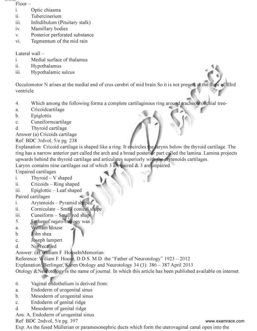
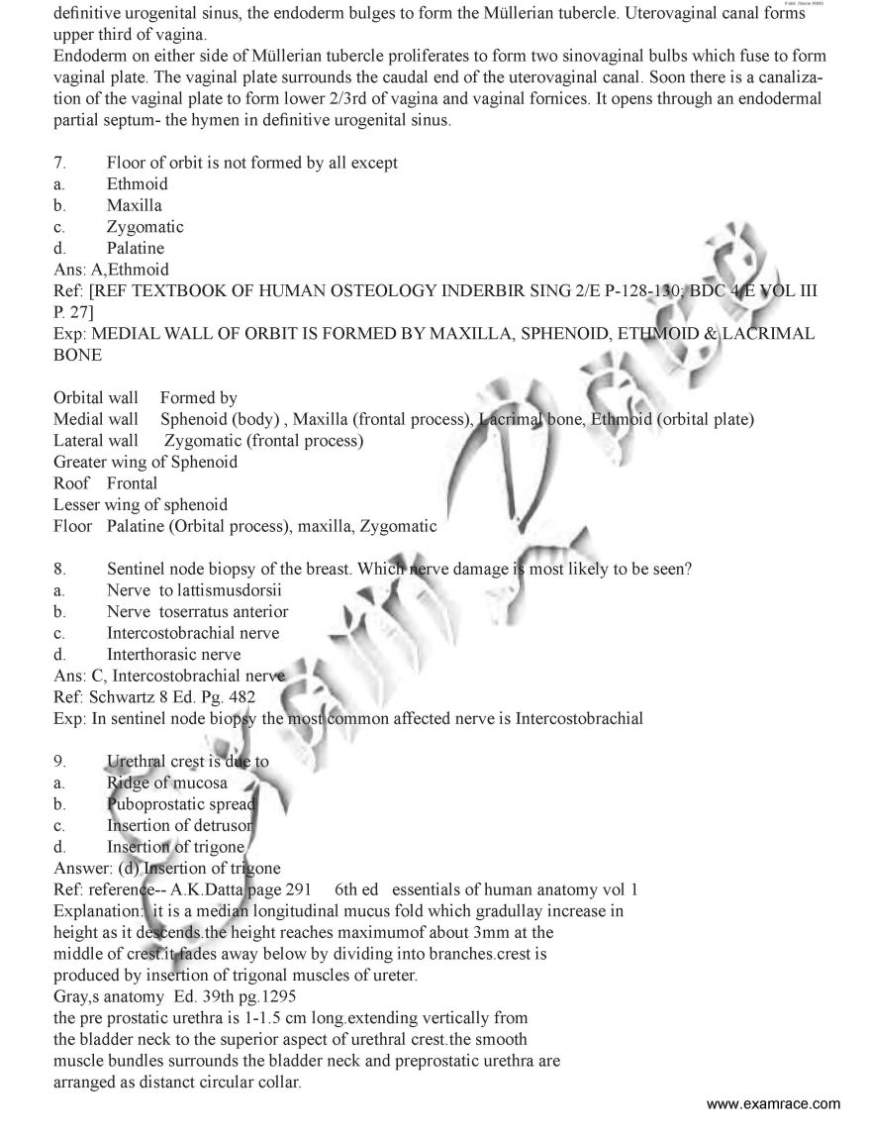
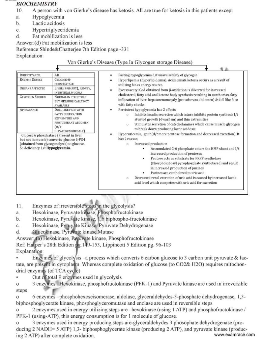
National Eligibility cum Entrance Test-Post Graduate is a national level examination conducted by National Board of Examinations for admission to MD/MS and PG Diploma courses in colleges across India. NEET PG entrance exam question paper ANATOMY 1. In the advancement of surgery of shoulder joint, one muscle was not given much importance earlier but now came in picture, this forgotten muscle of rotator cuff is a. Subscapularis b. Supraspinatus c. Infraspinatus d. Teres minor Answer: (a) Subscapularis Explanation:Musculotendinous cuff of shoulder is a fibrous sheath formed by the four flattened tendons which blend with the capsule of shoulder joint and strengthen it. Muscles which form the cuff arise from scapula and are inserted into the lesser and greater tubercles of humerus. They are subscapularis, supaspintaous, infraspinatus and teres minor. There tendons while crossing the shoulder joint become flattened and blend with each other on one end and with capsule of the joint on the other end, before reaching their points of insertion.Cuff gives strength to the capsule of shoulder joint all around except inferiorly. That is why dislocation of humerusoccur most commonly in a down ward insertion. 2. Stuctures at anorectal junction all except a. External sphincter b. Internal sphincter c. Anococcygeal raphe d. Puborectalis Answer (c)Anococcygealraphae Explanation: Anorectal ring is a muscular ring present at the anorectal junction. It is formed by the fusion of the puborectalis, uppermost fibers of external sphincter and internal sphincter. Surgical division of anorectal ring results in rectal incontinence. Anococcygeal ligament or anococcygeal raphe is a multilayer musculotendinous structure between anal canal & hip of coccyx. It is attached anteriorly to the superficial part of external anal sphincter i.e. middle part of external anal sphincter which lies below to deep part of external anal sphincter at the anorectal junction present deep part of external anal sphincter. 3. Structure not present at floor of third ventricle- a. Optic stalk b. Third nerve c. Infundibulum d. Mamillary body Answer: (b) Third nerve (Occulo motor N.) Explanation: Third ventricle is a median cleft b/w two thalami. Embryologically it represents the cavity of diencephalon. Communication :Antero superiorly, on each side it communicates with the lateral ventricle through the interventriclar foramen (or Foramen of Monro). Postero inferiorly it communicates with the fourth ventricle through the Cerebral Aqueduct. Boundaries: Anterior wall – i. Lamina terminalis ii. Anterior commissure iii. Anterior columns of fornix Posterior wall – i. Pineal body ii. Post commissure iii. Cerebral aqueduct Roof - i. Ependymal liningof the under surface of telachoroidea of third ventricle. Floor – i. Optic chiasma ii. Tubercinerium iii. Infndibulum (Pituitary stalk) iv. Mamillary bodies v. Posterior perforated substance vi. Tegmentum of the mid rain Lateral wall – i. Medial surface of thalamus ii. Hypothalamus iii. Hypothalamic sulcus      For more questions here is the attachment. |